Picture of the Week

Male and female pronuclei together
Sunday, June 6, 2010
This week’s picture is a follow-up to the previous post, which illustrated the products of meiosis in the nemertean worm Cerebratulus. Assuming that fertilization succeeds (i.e., one and only one sperm enters), and female meiosis completes, the egg finds itself inhabited by two haploid nuclei – one from the sperm, and one from the egg. These “pronuclei” need to get together to constitute the diploid nucleus of the zygote.
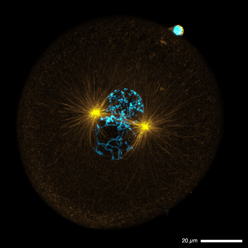
The word “zygote”, referring to the cell resulting from fusion of two gametes, apparently comes from a Greek root meaning “to yoke”, as in a team of animals. This etymology seems exceptionally fitting when considering the picture above, in which the microtubules radiating from the two centrosomes seem to embrace and tie together two as-yet-distinct orbs,* whose efforts must be synchronized to create a new animal.
In many species it appears that pronuclei find each other by the simple expedient that the male pronucleus is associated with the only microtubule organizing center in the cell (the maternal centrosomes having been discarded or destroyed during female meiosis). This single microtubule organizing center duplicates to become the two poles of the first mitotic spindle. Whether pronuclear migration happens before or after the sperm centrosome duplicates (and in Cerebratulus, it’s after), the female pronucleus associates avidly with the microtubules that grow from the centrosome. It slides along them, probably driven by the motor dynein, until it reaches the centrosome(s) where it finds the male pronucleus waiting.
There are several essential steps – pronuclear migration, duplication of the paternal centrosome, and DNA replication – but male and female pronuclei don’t necessarily fuse before first cleavage. In nematodes like C. elegans and in many molluscs, and probably other animals as well, the nuclei remain distinct and, at first mitosis, break down simultaneously to admit forming spindle fibers. In such cases the paternal and maternal genomes don’t cohabit the same subcellular compartment until after first cleavage. In contrast, in echinoderms the pronuclei fuse well before DNA replication. Cerebratulus must be somewhere in between; the chromosomes are clearly condensed in this picture, thus DNA replication must be complete. But other images in the sample from which this one was taken show a single, fused nucleus before formation of the mitotic spindle.
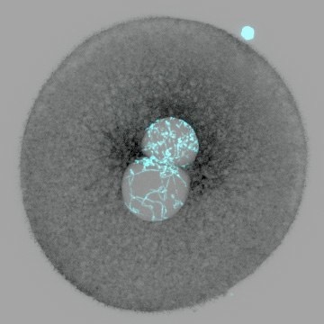
* It’s a little hard to appreciate, in the false-colored image above, that the male and female pronuclei remain distinct at this stage; here is a different representation of the same image data which makes this aspect more apparent:
Species:
Cerebratulus marginatus (nemertean)
What is it:
A fertilized egg in which male and female pronuclei have met
Points of interest:
Pronuclear fusion, centrosomes
What’s glowing:
Antibody to tubulin labels the microtubules (orange), Hoechst 33342 labels DNA (blue)
Optics:
Olympus FluoView 1000; 60x; projection of 91 0.3-µm sections.
Picture taken by:
George von Dassow
Please click the picture to see a full-size version (232 kb)


