
Pictures of Unusual Size
These pictures don’t lend themselves well to web slideshows because of their size. Each of the small versions below is linked to a large version that will open as a single image in the page, without the caption.
Several of these pictures were assembled from overlapping micrographs using ImageJ and the image stitching plugins written by Stephan Preibisch

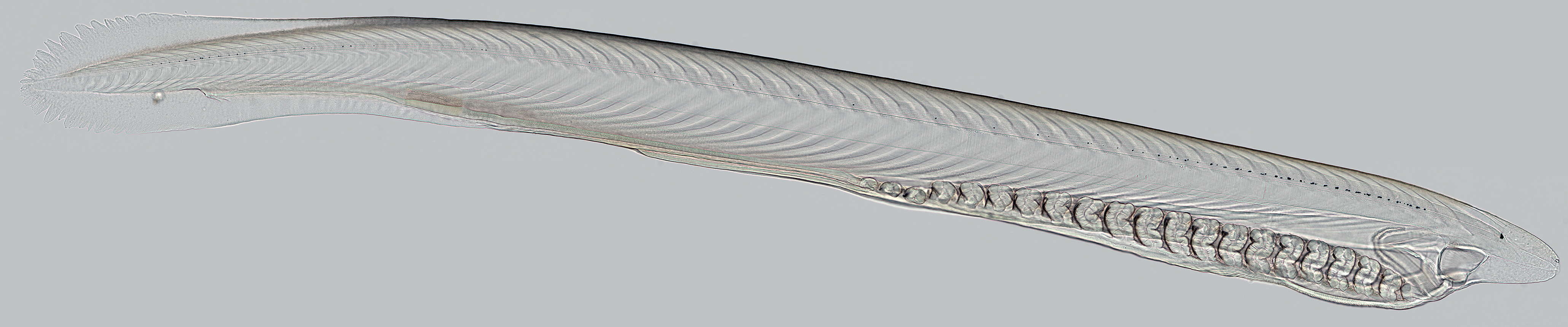
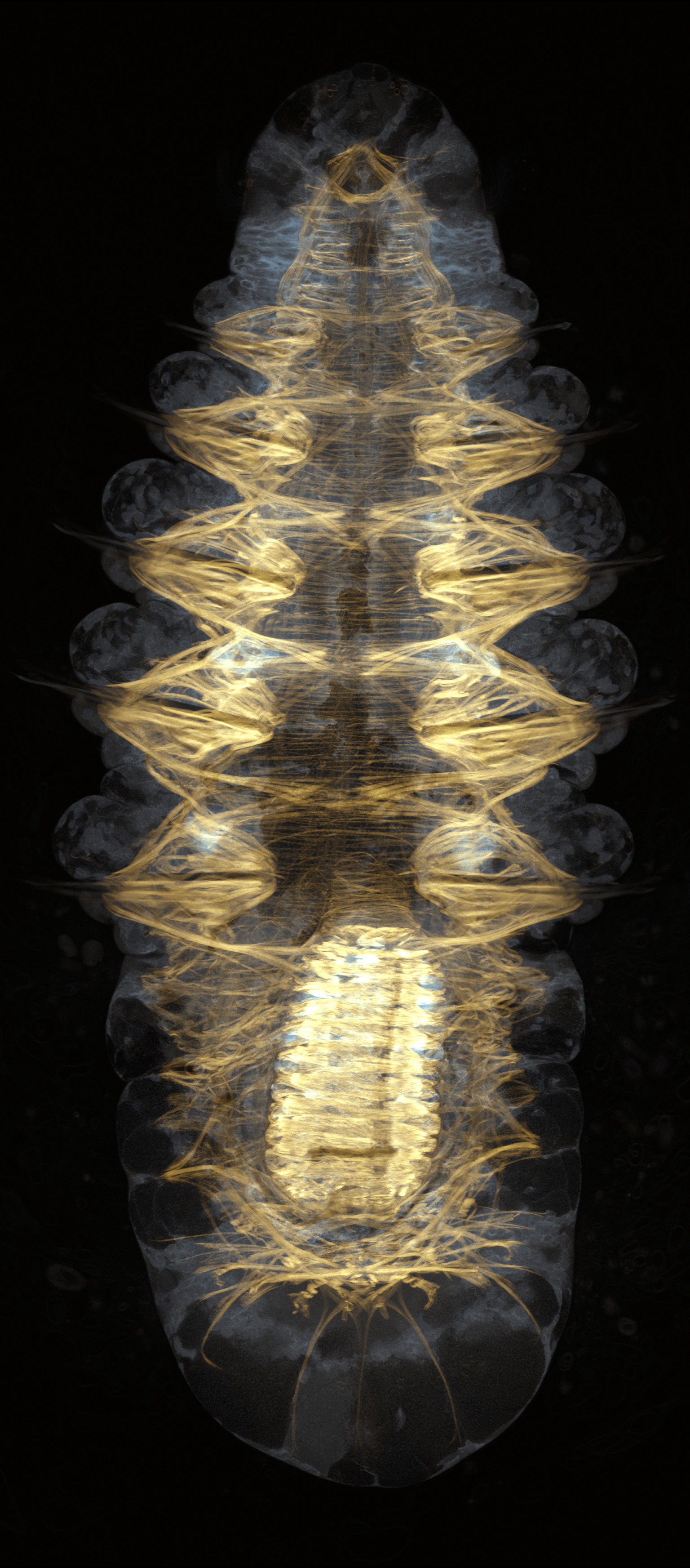
Montage of a larval lancelet from 19 overlapping pictures with a 20x lens. This animal was caught in surface plankton in the Gulf of Mexico, and held itself remarkably still and flat long enough for these pictures; I got from tip to tail without changing focus. The original fused image is >16,000 pixels wide, and clicking will open a 25%-reduced-but-still-large version.
Please remember, I made these images and they are mine. I don’t watermark them because it’s ugly and I would rather not have to do so. If you like them and find them useful, you are welcome to use them informally, as long as you specifically attribute them to me. Please do not display or print them elsewhere without my explicit permission.
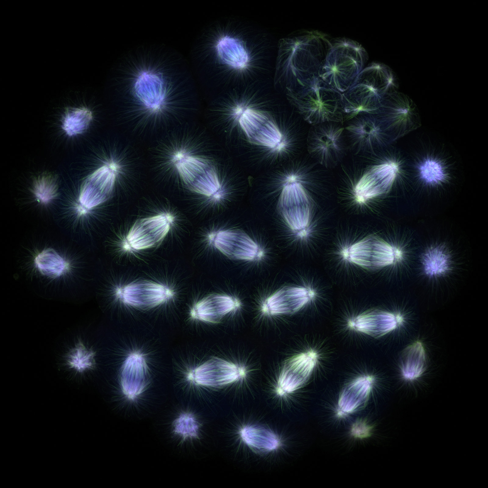
Projected focal series through the top quarter of a sand dollar blastula stained for tubulin, showing mitotic spindles. Small and non-mitotic cells to the northeast are progeny of the micromeres. The false coloring used the brightest-point projection for the green channel, and an average for the red and blue. The full size version is 1600 pixels square.
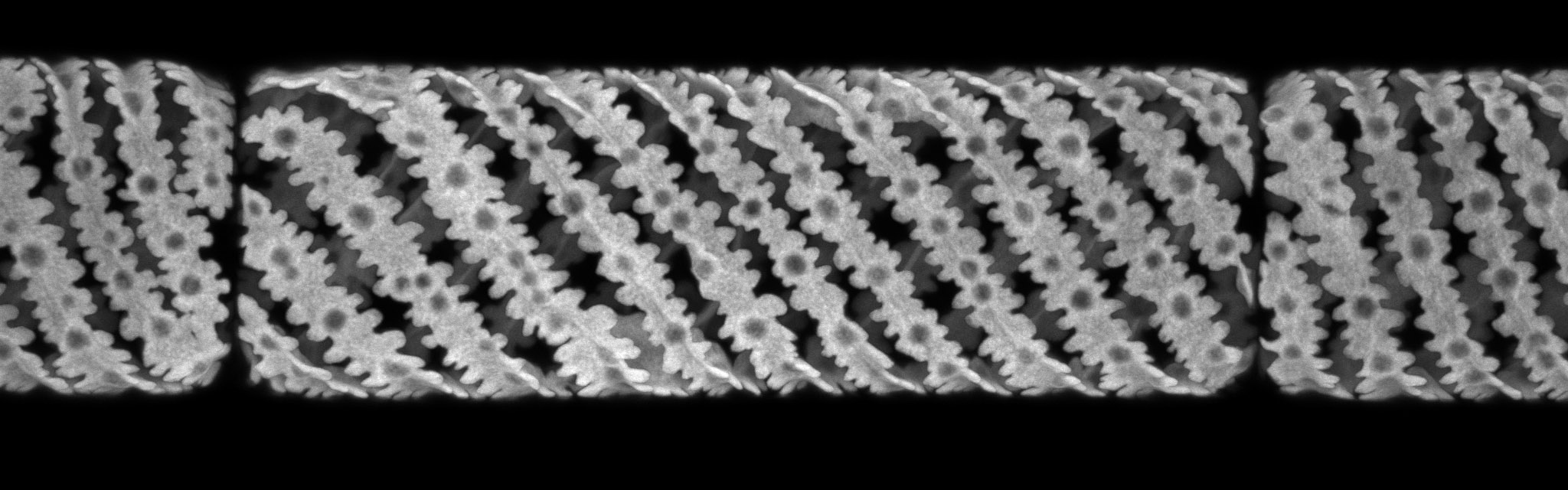
Projected focal series through part of a chain of diatoms (Stephanopyxis?). These cells are alive and unstained; this is the fluorescence of chlorophyll. The fusion is imperfect because the chloroplasts move around a bit. The full image is 2780 pixels wide.
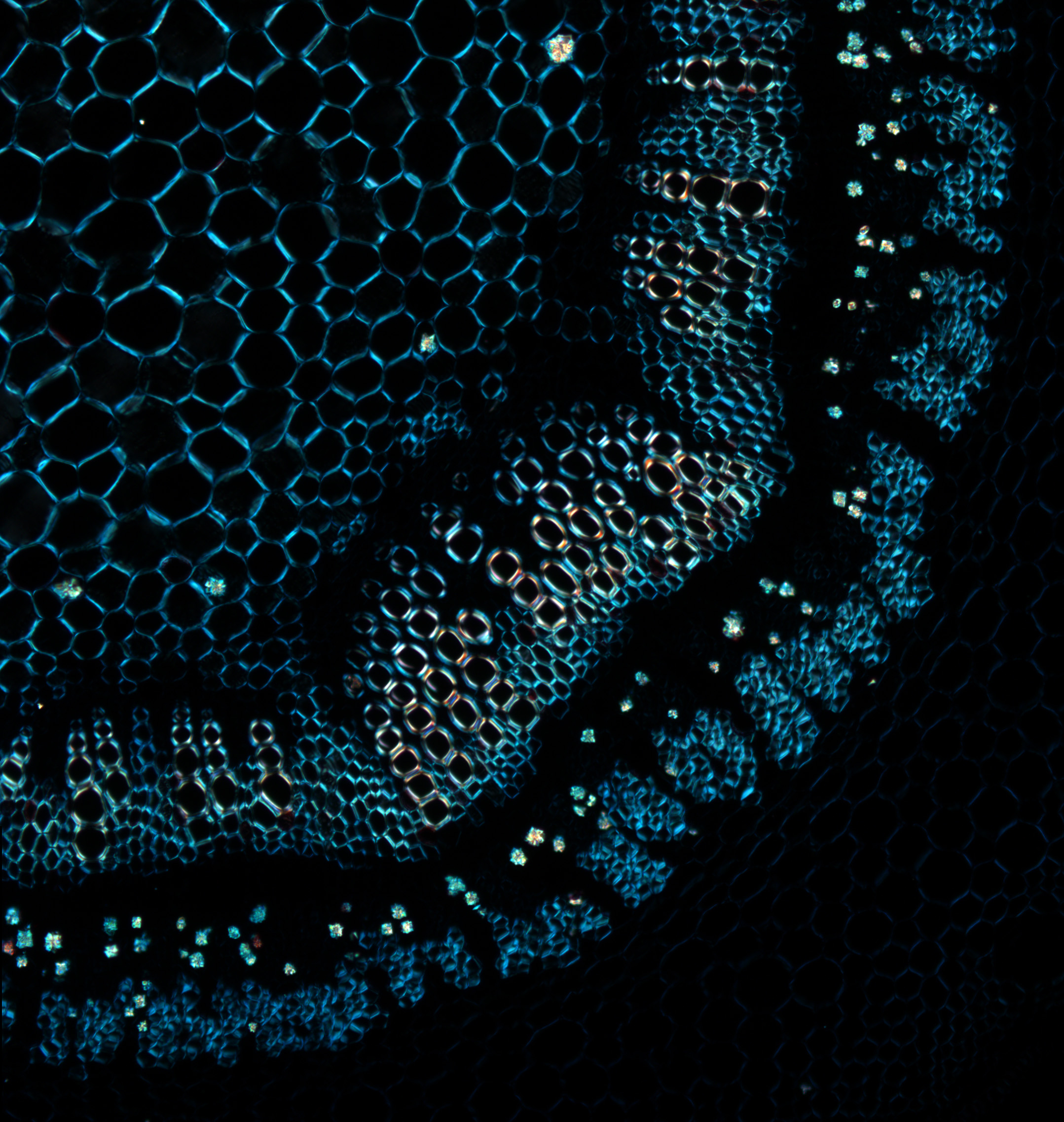
Part of a section of a cotton stem viewed in polarized light. The whole field of view in this montage is about 2 mm.

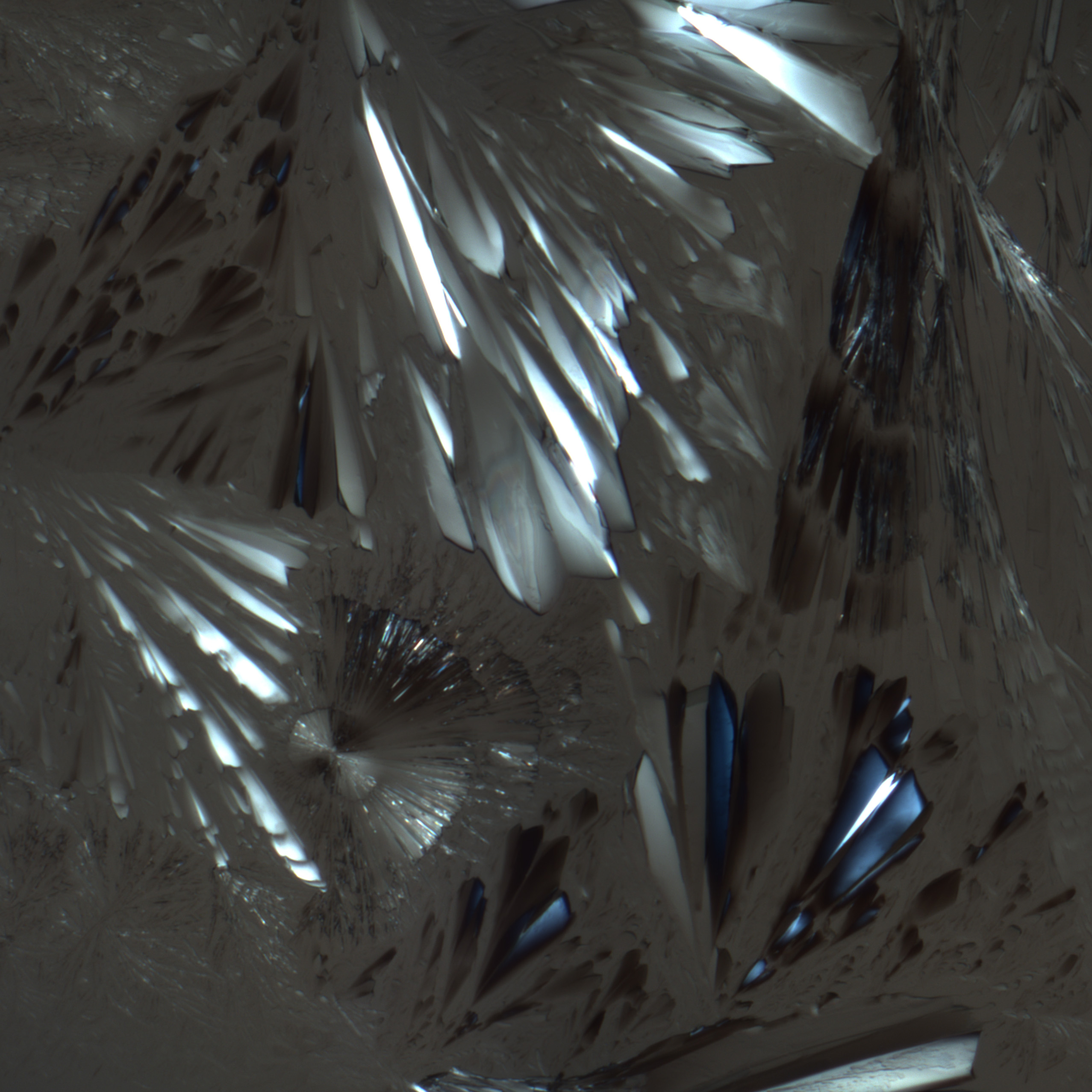
Mannitol crystals dried on a slide, viewed in polarized light, probably with a piece of cellophane stuck in the condenser. The large image is 2555 pixels square.
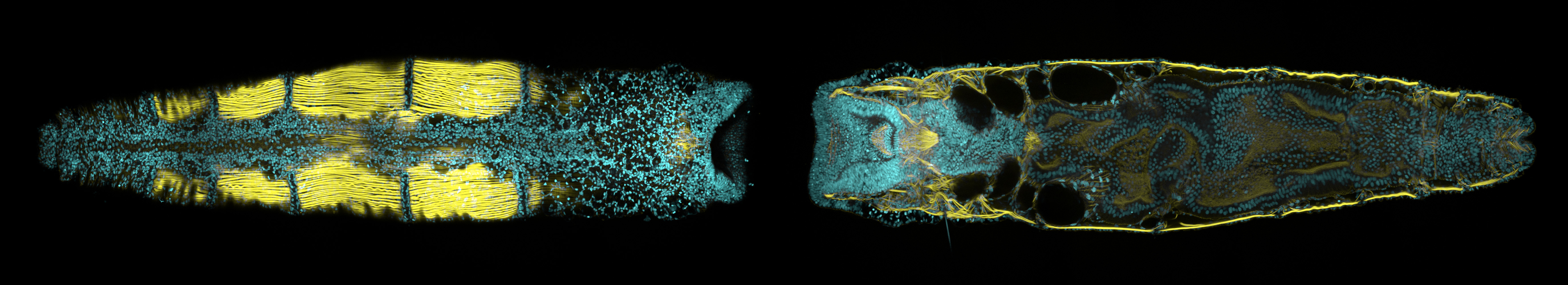
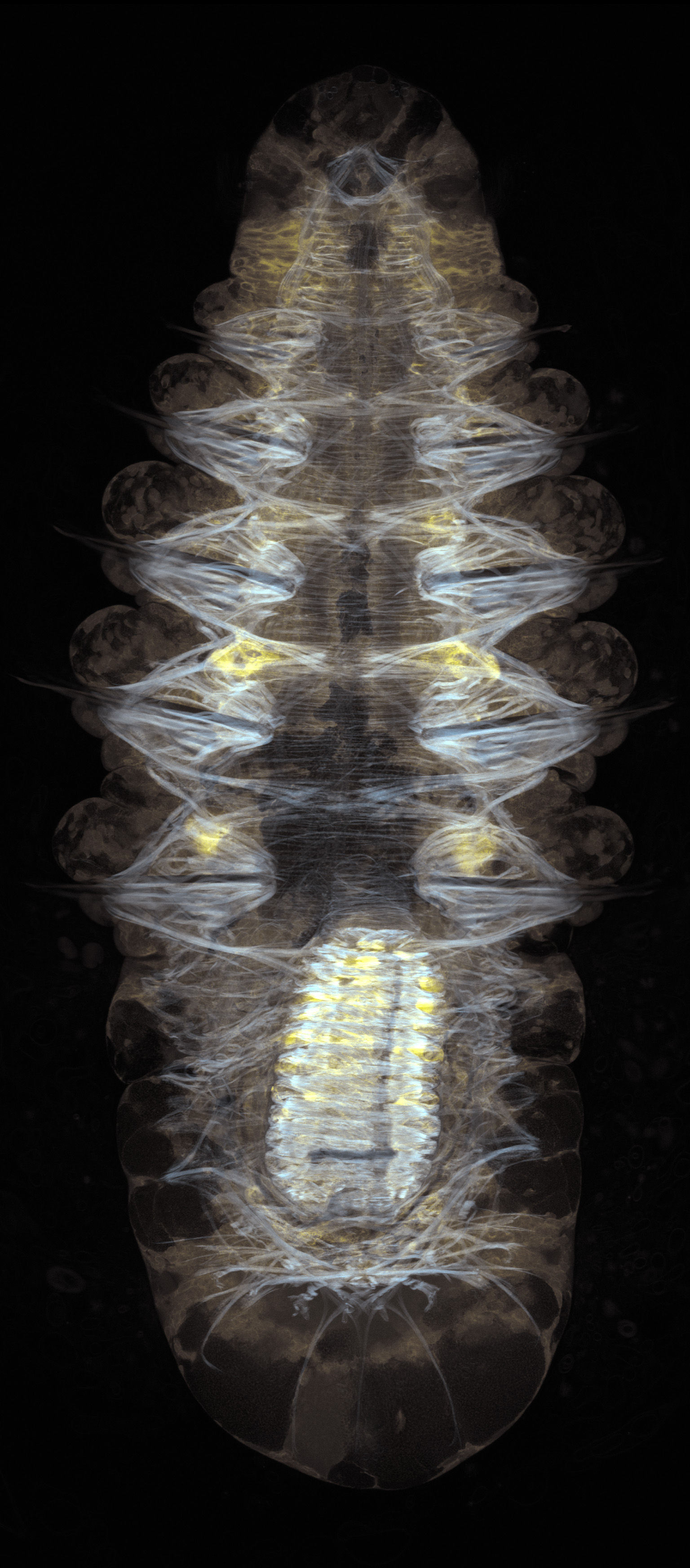
A juvenile Owenia collaris, at the surface and a frontal section. This individual was raised from egghood by Tracey Smart, and featured in our paper on Owenia’s development; I stained it with a DNA dye (blue) and phalloidin, which labels the muscles (yellow), and fused these together from overlapping confocal stacks. The full size image is 5500 pixels wide.

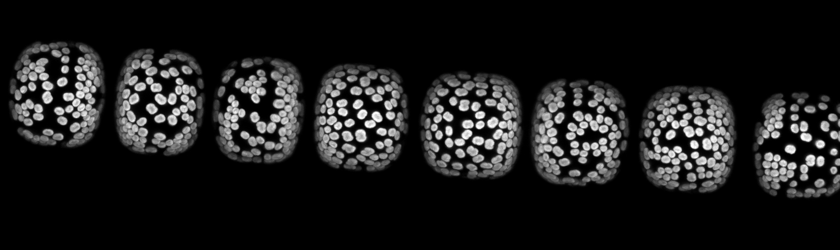
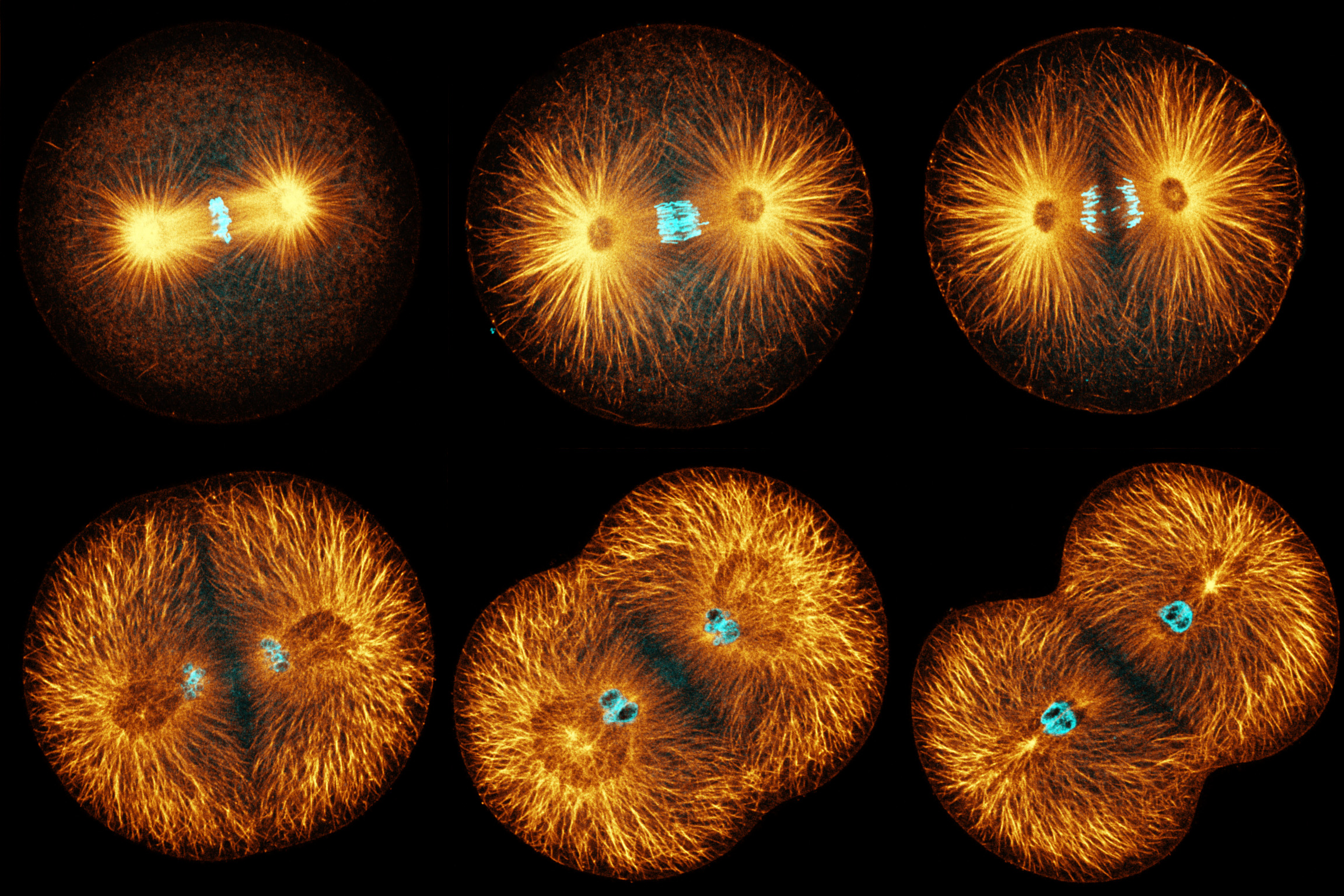
This is a matched set single confocal sections of sand dollar eggs at various stages of mitosis, stained for microtubules (orange) and DNA (blue); it was made as a figure for a paper I never wrote. The full image is 2304x1536.

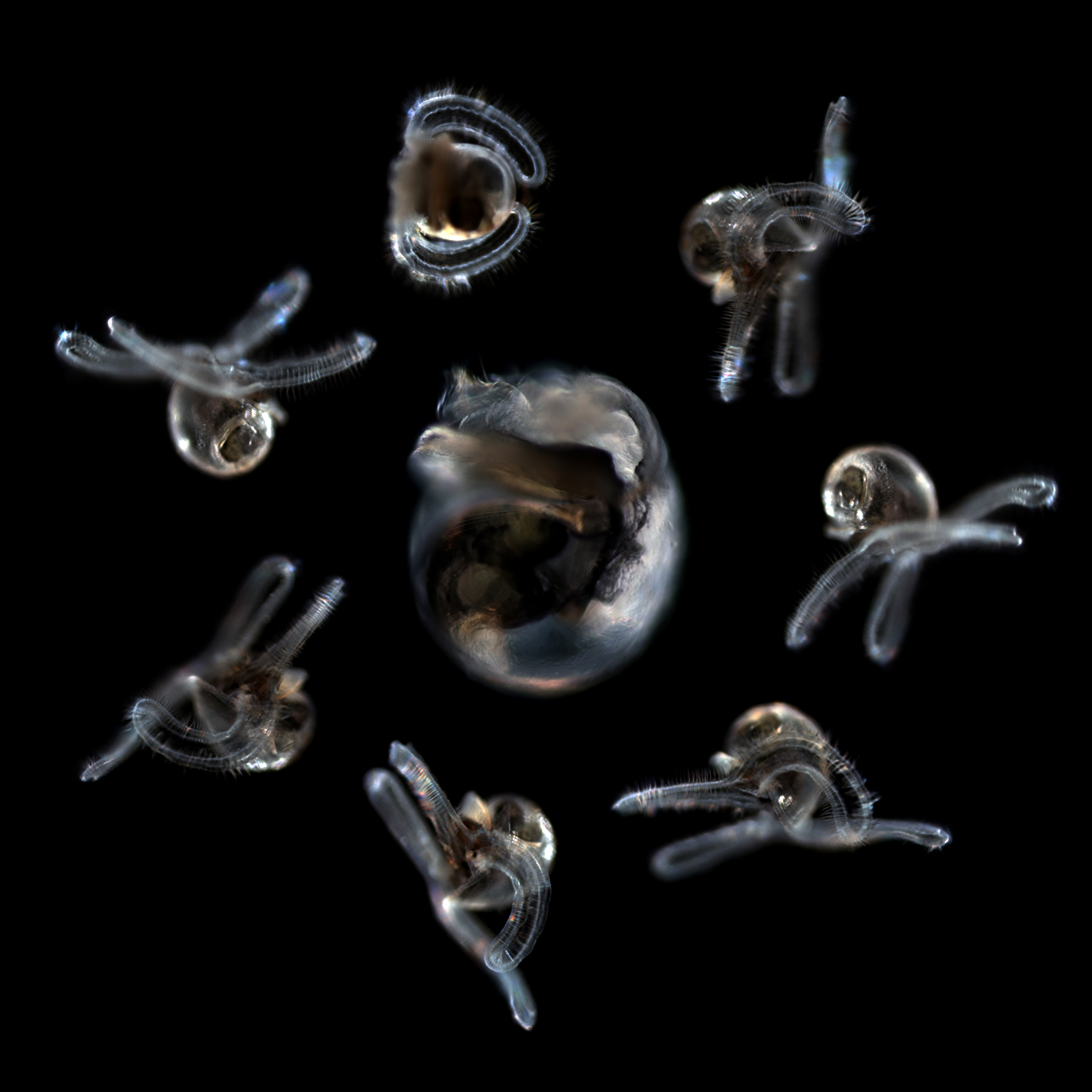
Darkfield photos of the same large veliger, caught in the surface plankton in the Gulf of Mexico. It may be the almost-fully-grown larva of Bathynerita, a snail that lives around cold seeps. The montage was made for an auction to benefit the Strathmann Fund. The full image is 2596 pixels square.
Projected confocal series through a filament of Spirgyra of some sort; these are living cells, revealed by the fluorescence of chlorophyll. The full size image is 2048 x 512.
